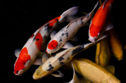
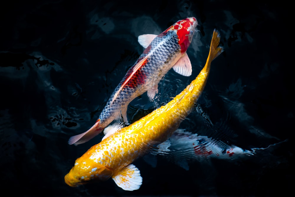
Many people keep Koi as pets and as landscaping “gems” in their ponds or water fountains.
It’s no wonder koi fish are frequently connected with character strength, tenacity, success, and courage. The fish also represents luck, success, prosperity, and ambition. Koi are also associated with longevity due to their long history and tenacious nature.
Did you know that your koi and pond fish have a two-chambered heart? Should that even matter to you as the owner of a fish-filled pond? Most likely not; however, you should know koi and pond fish anatomy if it is important to you.
Read on if you’re interested in learning more about these incredible fish!
Anatomy of the Koi Fish
External Anatomy
Fins
Fins – Koi have three single fins and two pairs of paired fins. The caudal fin, often known as the tail fin, is largely utilized for forward swimming, particularly rapid swimming. The dorsal (top) fin is employed for forward motion stability. As the Koi grows older, the biggest leading ray stiffens, sharpens, and turns thorn-like.
The anal fin, like the dorsal, serves as a stabilizing mechanism. In addition, when the Koi grow older, the biggest leading ray stiffens, sharpens, and becomes thorn-like. Finally, the pectoral fins are paired and serve various purposes, including guiding during forward motion, slow swimming forward and backward, breaking, and counteracting the jet effect caused by water being driven out of the opercular apertures.
The pelvic (or ventral) fins are paired and help to regulate pitch and roll as well as counter lift. The fins are slender and strongly vascularized, making them prone to injury. Changes and damage to the fins are also easily visible. As a result, infections are frequently identified in the fins, which might seem injured, torn, or bleeding.
Cuticle
The cuticle, a non-cellular mucous layer, protects the skin and scales. The cuticle of the Koi, also known as the slime coat, is a thin coating of mucus that includes several defensive compounds like antibodies, lysozyme (an enzyme that is damaging to the cell walls of some bacteria), and C-reactive protein (a protein that may have some antibacterial properties).
The cuticle is the Koi’s first line of defense against waterborne irritants and parasites, and it helps the skin reduce drag for improved movement.
Skin
The skin’s cellular layers are epidermis, dermis, and hypodermis. The epidermis (outermost cellular layer) is thin, often just 6 to 8 cells thick, and includes unicellular mucous glands connected by a network of tiny capillaries.
The scales, scale-forming cells, pigment, blood arteries, and nerves are all found in the dermis (middle layer). The hypodermis is a vascularized fatty layer between the epidermis and the muscle or bone underneath it. It serves as the link between the skin and the rest of the body.
The skin of a swordtail (tropical livebearer) is depicted below; however, except for the contour shape of the scales (seen later in this section), it is quite similar to carp skin (Koi)
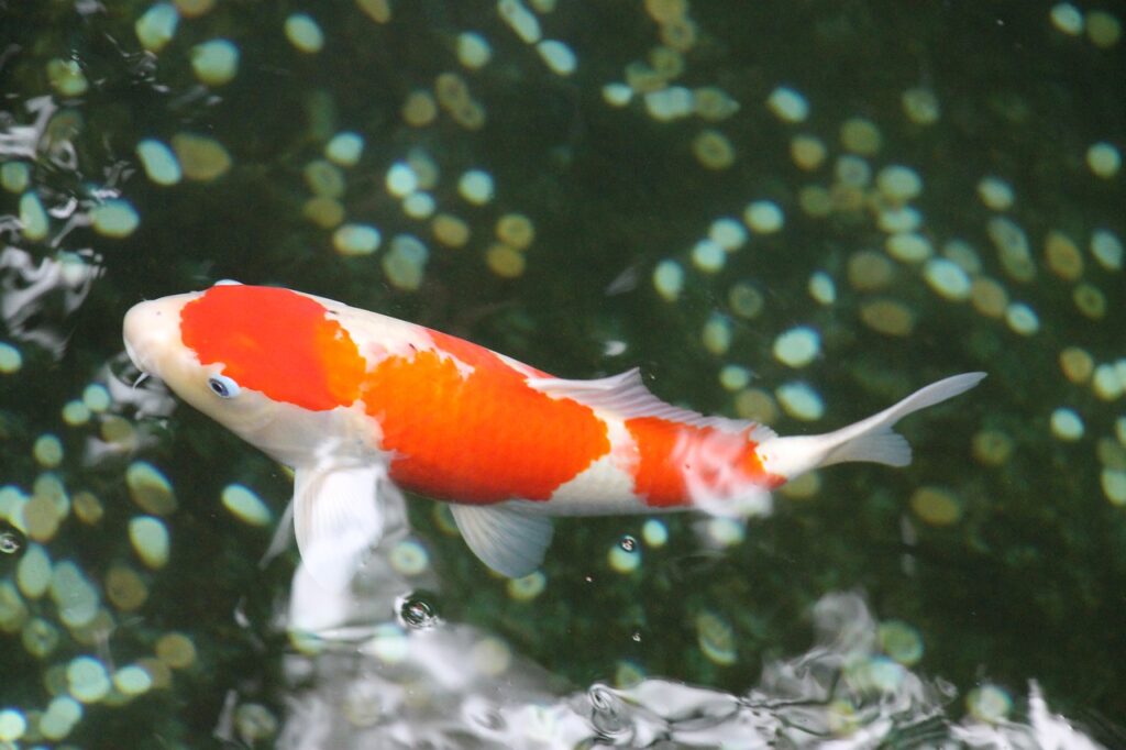
Scales
All koi scales are cycloid, and most Koi have scales on most of their bodies but not their heads. In Japanese, these Koi sized Koi are known as wagoi.
Some koi have scales exclusively on the dorsal and lateral lines, known as German scaled Koi (Doitsu, in Japanese). Some koi/carp have no scales and are known as leather koi/carp. Other koi/carp have massive scales that arise nearly randomly and are known as armored scaled koi/carp.
Scales are layered, thin, flexible plates that emerge from the dermis. Because the scales are formed in the dermal layer, removing a scale generates an ulcer, or hole in the skin, which can be a possible entry point for infections. The scales expand mostly from the middle outward.
There is a lot of overlap in the scales, and there can be up to six or seven levels in certain places (small dark areas). However, approximately 20% of most scales are exposed to the outside, i.e., the region of the scale without overlapping scales, as shown in the huge dark area in the figure above.
The dermis develops from behind the scale on certain Koi and is known as ‘fukurin.’ Fukurin is commonly found in ogon and asagi, particularly on the shoulder. It can be found on the scales’ outer edges. The fukurin is the platinum or white spots on the main body of the fish depicted on the right, whereas the scales are the darker or yellowish parts.
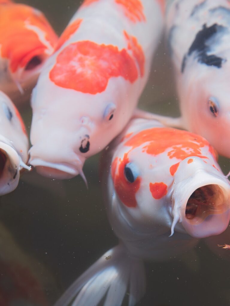
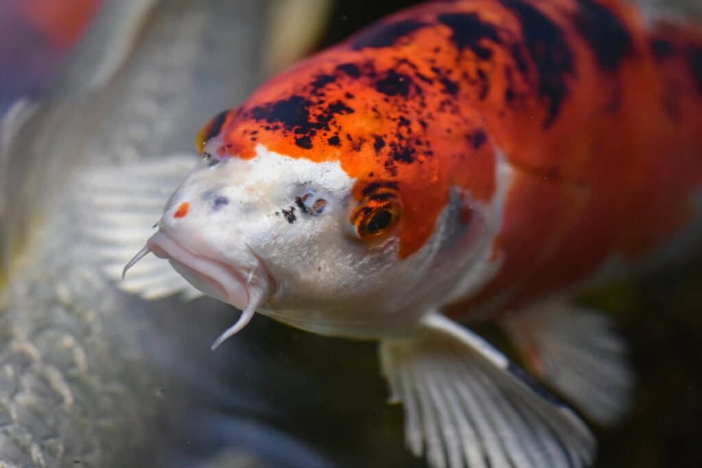
Mouth
Koi have a protruding mouth that is perfect for bottom feeding. Because the location of the mouth on the underside of the head allows them to suck meals off the pond bottom physically, Koi are known as benthic feeders. They may eat at any depth, including the bottom, mid-water, and top.
Koi have no teeth in their mouths, which is compatible with how Koi feed in the wild; that is, they suck everything up, taste it, and then decide if it is edible or not. The Koi, on the other hand, have tooth-like features (pharyngeal teeth) on their pharyngeal bones, which are placed directly beneath the gill chambers. These teeth grind food against a bony pad (carp stone) placed on the roof of the throat.
The tail
The tail fin of your koi fish is divided into five sections: the base, the middle region, the upper portion, the lower portion, and the tip. This is the starting point for the full length of the tail fin. It’s typically half the size of the remainder of the tail fin.
The middle section begins above the base and continues down to the tail fin. It is usually double the width of the base. The upper part of the tail fin is located just above the middle region. This part is larger than the middle section.
The bottom portion of the tail fin, which is thinner than the central segment, is located underneath the center section. Therefore, the site of connection to the body is located near the bottom of this section.
The tip is located at the tail’s end. It has a wedge shape and is frequently colored differently from the rest of the tail. Spots or stripes may appear on some types.
Organs of Sensation
Hearing
Koi do not have external ears but instead, hear through internal “ears” linked to the cerebral swim bladder via a collection of bones known as the Weberian ossicles. It is thought that the swim bladder amplifies sound.
Koi, like other fish, are particularly sensitive to sound and can get agitated to the point of being unwell if repeatedly exposed to loud noises. The figure below depicts the sensory systems of sea trout heads, but it is thought to be typical of the concept and layout of koi/carp inner ears.
Eyes
The anatomy of koi eyes is similar to that of our own. They do, however, have bilaterally located eyes that can move independently, which widens the range of area in their vision field. In addition, they have cones and rods, which are the structures of the eye that see color, black and white, and grayscale, respectively.
They most likely have decent enough vision to recognize the form of words on a printed page. In addition, Koi do not require protective eyelids because they live in water.
This, however, increases the risk of abrasion or other mechanical stress to their eyes during netting and handling.
The lenses are spherical and protrude through the pupil, allowing for extensive peripheral vision, including front and back. The lens of a healthy fish is entirely clear. However, bacterial and nutritional issues can cause the lens to fog. The pupils are controlled neurologically in the same way as mammals are, but the reaction time is too sluggish for therapeutic application.
The Lateral Line
The lateral line runs along the side of the Koi, approximately halfway down the fish. Holes in the scales connect to a canal beneath the surface containing neuromast cells. Any movement of water touching the sides of the fish causes the mucus in the canal to vibrate.
These vibrations excite the neuromast cells, which are related to the peripheral nervous system, giving one of the most powerful survival senses (flight reaction). The lateral line is a crucial marker. It is at the same level as the spine, which has a blood artery that runs along the length of the spine immediately ventral (underneath) to the spine and will be useful in detecting the blood vessel.
Olfaction
The olfactory organs (used to smell) are positioned near the base of the nostrils, known as the nares. Water does not pass from the nares to any other body portion. They are paired and placed between the eyes and the mouth; they are solely utilized for olfaction (smelling).
They are fashioned like miniature U-tubes into which water enters via the leading or forward hole and escapes through the back port or aperture. As the Koi travels forward in the water, a flap of skin just below the front aperture sends water into the forward entrance of the nare.
Diffusion and the movement of little hair-like structures (cilia) within the nares help transport substances via the nares.
Taste – Barbels
Taste buds are abundant in and around the lips, mouth, and barbels. The barbels of Koi are divided into two pairs. They had three couples three hundred years ago.
Bones
Carp are Teleost fish, which means “bony skeleton,” among the boniest freshwater fishes. Fish bones are light and thin, having no bone marrow in the core. A lightweight skeleton is helpful to an animal that must be buoyant to survive in an aquatic environment.
Muscle
Teleost fish have two types of muscle: white muscle and black muscle. The white muscle is supposed to be for fast, swift swimming, whereas the black muscle, placed just beneath the skin, is thought to be for slow, steady swimming. The clinical importance of these diverse kinds of muscle is that when drugs are injected into them, they have a wide range of drug kinetics.
It is widely assumed that red muscle is well vascularized (has a good blood supply), which would facilitate absorption of medicines into the blood faster than white muscle and sustain the release because the blood flow is also supported, as opposed to white muscle, which can create an oxygen debt and requires a recovery period.
Gills
The gills are a complicated system of rigid bony and cartilaginous arches. The fish has four gill arches on each side. The gill filaments are located outside the gill arch and are made up of lamellae and secondary lamellae structures, the latter of which is only 2 thick.
These structures are in charge of exchanging gases (O 2 / CO 2) across the epithelial cells. The epithelial cells are a direct conduit between the oxygen in the water and the fish’s bloodstream.
Mucus-producing and chloride cells, which serve in mucus formation and osmoregulation, are also found in the gills. The operculum, also known as the gill cover, is a structure that covers the gill arches on the exterior of the fish.
The principal function of this multi-purpose portion of the Koi’s anatomy is to assist manage the pressure of the water taken in by the mouth and transmitted through the gill filaments. Gill rakers, which operate as food filters, are also located on the gill arches but in the inner or anterior section of the arch. Clogged rakers are frequently the source of flashing or shaking when the fish tries to clean the gills of extra food after feeding.
Internal anatomy of Koi
System of Circulation
The heart is the pump that circulates blood throughout the body. It is found towards the bottom of the fish, between the gills. The heart of a koi is a two-chamber organ with a ventricle and an atrium. Although it is a four-chambered organ, the extra two chambers before and after the main pumping chambers are known as the sinus venosus and bulbus arteriosus, respectively.
They are smaller than the main pumping chambers and serve as accumulators (to level out pressure spikes and protect the cardiovascular system from overpressure) rather than pumping blood. They lack muscular barriers yet are elastic, much like balloons. As a result, fish have far lower blood pressure than mammals.
Blood goes from the heart to the gills, disseminated throughout the body. Next, blood is gathered into veins, which finally return to the sinus venosus just before returning to the heart. In contrast to mammals, fish have just one circulation pattern.
Mammals have both systemic and pulmonary circulation. The caudal vein, one of the most important veins, is ventral (under) the spine. This is the easiest vein in the fish to get blood samples from.
Normally, blood goes from an artery to an arteriole, then to a capillary, then to a venule, then to a vein, and finally back to the heart. A portal system is one in which blood passes from a vein into a capillary and then returns to the vein on its journey to the heart. For example, the liver circulates through a portal in fish and humans. In other words, blood from the intestines passes through liver capillaries before collecting in the hepatic portal vein.
Fish have a renal portal system as well. However, Carp (Koi) has a somewhat different system than the standard. Blood returning through the caudal and segmental veins in carp is separated into the renal portal vein and a shunt to the intestinal vein.
Kidneys
In a koi, there are two kidneys. The caudal kidney is a long and thin organ that runs almost the length of the body cavity and is placed right below the spine. The anterior, head or cranial kidney is the other. It is placed right above the heart and contains Thyroid follicles as well.
Liver
The liver is smooth and dark red-brown and is located near the cranial section of the intestine. The gallbladder is protected by the right lobe of the liver, whereas the left lobe protects the spleen.
Bladder for swimming
This is a two-chambered organ positioned exactly beneath the kidney and directly beneath the spine. Between the two chambers, there is a short link that also connects to the stomach. The most caudal chamber is relatively rigid, although the other chamber has some wiggle room.
Gut
Mechanical food breakdown and mixing begin with the pharyngeal teeth, followed by chemical breakdown and absorption in the long intestine. The leftover solid/undigestable material is discarded after the nutrients have been absorbed. Therefore, Koi are not able to digest food.
When Koi have a full gut during the winter, the lining may get compromised, allowing dangerous germs to pass through and enter the bloodstream. Unfortunately, they may not exhibit any signs of this until the weather warms up in the spring.
Gonads
The gonads are found between the swim bladder and the intestines. They can be separated or combined. The ovaries are pink and smooth, whereas the testes are white and fissured. The gonadal pore is distinct from the waste pores at the anal vent. During the mating season, gonads grow to about 70% of their body weight in females and around 30% in males. Male and female gonadal tissues can be found in certain Koi. These hermaphrodites have male and female outward traits.
FAQs
Is it true that koi fish have nostrils?
The fish are a subspecies of the common carp, Cyprinus carpio, and comprise a variety of wild carp races and cultured Koi (“Nishikigoi”).
The resultant fish featured larger fins, longer barbells, and pom pom nostrils, as well as being tougher than Koi.
Koi fish have how many fins?
Fins. Koi have two pairs of paired fins (the pectoral and ventral, or pelvic) and three single fins (the caudal, or tail, dorsal and anal fins).
What causes Koi fish to open their mouths?
Your pond’s water most likely lacks enough dissolved oxygen for the fish to “breathe”; thus, they’re gasping for oxygen from the air due to a lack of aeration or poor water quality. To collect dissolved oxygen from the pond, Koi breathe by pushing water over their gills.
Is it true that koi fish have teeth?
Koi carp have teeth that they use to eat their food. However, they don’t have them in their mouths! This is because the koi carp’s teeth are located deep back in their throats, rather than in their mouths, so when they take food into their mouth, it is chewed and digested via the throat before entering the stomach.